
The specimen images below are source-attributed photographs. Editorial source matching is not independent expert identification. Credit and licence information applies to the individual image, not to the rest of this site.
CDC-origin material is freely available from the CDC Public Health Image Library (new tab). Inclusion does not imply endorsement by CDC or HHS. Some displayed images are adaptations described below.
Adult bed bug in profile
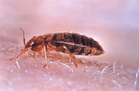
Adult bed bug in profile. Photo: CDC / World Health Organization, Geneva. Public domain. Source: CDC PHIL; original image available free from CDC. Use does not imply CDC, HHS, or U.S. Government endorsement.
Adult bed bug in profile.
Profile view of a feeding specimen; use the dorsal image for overall outline. This photograph cannot identify the cause of a skin mark or distinguish every Cimex species.
Original source (new tab) · Public domain
Changes: Resized and encoded as WebP; composition and substantive visual content preserved. Derivative image licence: Public domain.
Female German cockroach with an egg case, underside view
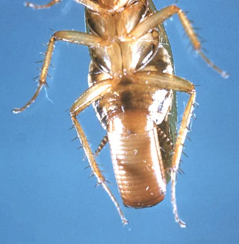
Female German cockroach with an egg case, underside view. Photo: CDC. Public domain. Source: CDC PHIL; original image available free from CDC. Use does not imply CDC, HHS, or U.S. Government endorsement.
Female German cockroach with an egg case, underside view.
Underside view with the head partly outside the frame. It illustrates the carried egg case; it does not show the two dorsal pronotal stripes used for recognition.
Original source (new tab) · Public domain
Changes: Resized and encoded as WebP; composition and substantive visual content preserved. Derivative image licence: Public domain.
Indianmeal moth at rest on a surface
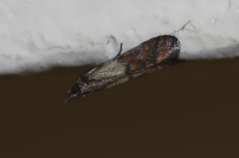
Indianmeal moth at rest on a surface. Photo: Fvukajlovic. CC BY 4.0.
Indianmeal moth at rest on a surface.
A single adult with folded wings. Lighting changes apparent color; confirm two-toned forewings and inspect stored food for larvae and webbing.
Original source (new tab) · CC BY 4.0
Changes: Resized and encoded as WebP; composition and substantive visual content preserved. Derivative image licence: CC BY 4.0.
Adult red flour beetle
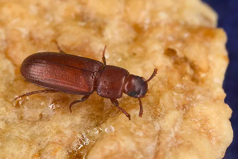
Adult red flour beetle. Photo: Peggy Greb, USDA Agricultural Research Service. Public domain. Use does not imply USDA ARS endorsement.
Adult red flour beetle.
A magnified example of one pantry beetle. Fine antennal characters distinguish it from the confused flour beetle; other stored-product beetles require separate identification.
Original source (new tab) · Public domain
Changes: Resized and encoded as WebP; composition and substantive visual content preserved. Derivative image licence: Public domain.
House mouse photographed in Ottawa, Canada
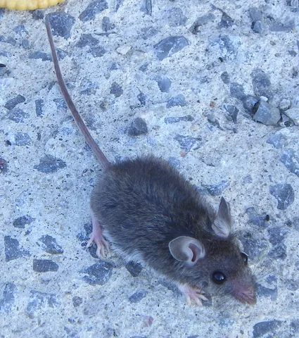
House mouse photographed in Ottawa, Canada. Photo: Ahunt. CC0.
House mouse photographed in Ottawa, Canada.
One adult photographed from above. No scale is present; apparent image size cannot distinguish a mouse from a young rat. Confirm body proportions and other evidence.
Original source (new tab) · CC0
Changes: Resized and encoded as WebP; composition and substantive visual content preserved. Derivative image licence: CC0.
Norway rat at Hatchpond, Poole, England
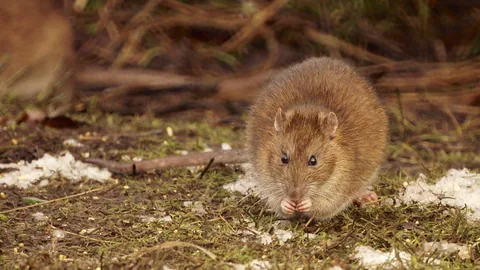
Norway rat at Hatchpond, Poole, England. Photo: Ian Kirk. CC BY 2.0.
Norway rat at Hatchpond, Poole, England.
Front view hides the tail and much of the body. It is an example of a rat, not a complete rat-versus-mouse or Norway-versus-roof-rat identification plate.
Original source (new tab) · CC BY 2.0
Changes: Resized and encoded as WebP; composition and substantive visual content preserved. Derivative image licence: CC BY 2.0.
Adult female lone star tick
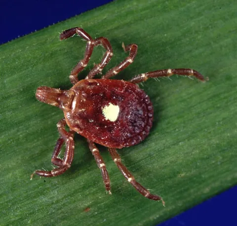
Adult female lone star tick. Photo: James Gathany; CDC / Michael L. Levin. Public domain. Source: CDC PHIL; original image available free from CDC. Use does not imply CDC, HHS, or U.S. Government endorsement.
Adult female lone star tick.
One adult female. Males, nymphs, larvae, and engorged ticks differ. No photograph determines whether a tick carries a pathogen or whether transmission occurred.
Original source (new tab) · Public domain
Changes: Resized and encoded as WebP; composition and substantive visual content preserved. Wikimedia source had already removed a small border; no further crop made. Derivative image licence: Public domain.
Female cat flea, preserved ZSM collection specimen
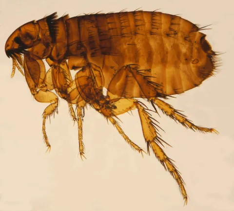
Female cat flea, preserved ZSM collection specimen. Photo: Katja ZSM. CC BY-SA 3.0.
Female cat flea, preserved ZSM collection specimen.
Magnified preserved specimen in side view. Color and posture differ from a live flea. Flea species identification needs fine characters; this cannot diagnose an animal or a skin mark.
Original source (new tab) · CC BY-SA 3.0
Changes: Resized and encoded as WebP; composition and substantive visual content preserved. Derivative image licence: CC BY-SA 3.0.
Winged termite photographed in Maun, Botswana
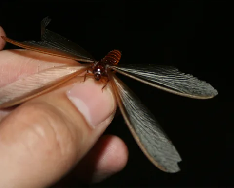
Winged termite photographed in Maun, Botswana. Photo: Discott. CC BY-SA 3.0.
Winged termite photographed in Maun, Botswana.
Species unspecified and photographed in Botswana. Use only as a generic example of termite wings, never as a named North American termite. Holding insects is not recommended identification practice.
Original source (new tab) · CC BY-SA 3.0
Changes: Resized and encoded as WebP; composition and substantive visual content preserved. Derivative image licence: CC BY-SA 3.0.
Winged male ant during a swarm
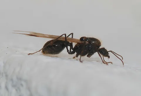
Winged male ant during a swarm. Photo: Alvesgaspar. CC BY-SA 3.0.
Winged male ant during a swarm.
A male ant with no species identification. Male antennae can look straighter than a worker or queen; do not use antenna angle alone to separate ants from termites.
Original source (new tab) · CC BY-SA 3.0
Changes: Resized and encoded as WebP; composition and substantive visual content preserved. Derivative image licence: CC BY-SA 3.0.
Pea aphids on an alfalfa leaf
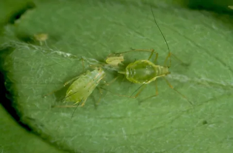
Pea aphids on an alfalfa leaf. Photo: Frank Peairs, Colorado State University / Bugwood.org. CC BY 3.0 US.
Pea aphids on an alfalfa leaf.
One aphid species on alfalfa. Other aphids vary in color and body form; host and close examination matter. The photograph does not set a treatment threshold.
Original source (new tab) · CC BY 3.0 US
Changes: Resized and encoded as WebP; composition and substantive visual content preserved. Wikimedia source history records watermark removal; the full required credit is retained here. Derivative image licence: CC BY 3.0 US.
Tobacco hornworm on a jalapeño pepper plant
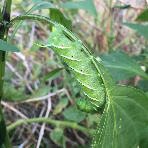
Tobacco hornworm on a jalapeño pepper plant. Photo: Macgeek13. CC BY-SA 4.0.
Tobacco hornworm on a jalapeño pepper plant.
A tobacco hornworm example. The rear horn is not clearly shown in this view; do not relabel it as a tomato hornworm or use it alone to separate the two species.
Original source (new tab) · CC BY-SA 4.0
Changes: Resized and encoded as WebP; composition and substantive visual content preserved. Derivative image licence: CC BY-SA 4.0.
Norway rat museum specimen LM1004, side view
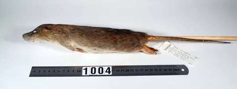
Norway rat museum specimen LM1004, side view. Photo: Auckland Museum. CC BY 4.0.
Norway rat museum specimen LM1004, side view.
Preserved museum specimen, not a living animal. Preparation changes posture and shape; the tail reaches beyond the photograph edge. Do not calculate a tail-to-body ratio from this image.
Original source (new tab) · CC BY 4.0
Changes: Resized and encoded as WebP; composition and substantive visual content preserved. Derivative image licence: CC BY 4.0.
Adult German cockroach, color dorsal view
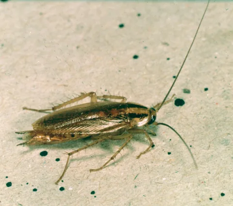
Adult German cockroach, color dorsal view. Photo: Clemson University - USDA Cooperative Extension Slide Series / Bugwood.org. CC BY 3.0 US.
Adult German cockroach, color dorsal view.
Adult dorsal view. Similar-looking Asian cockroaches require behavior, habitat, and potentially expert examination; color and stripes alone are insufficient.
Original source (new tab) · CC BY 3.0 US
Changes: Resized and encoded as WebP; composition and substantive visual content preserved. Wikimedia source history records a crop of its watermark; the full required credit is retained here. Derivative image licence: CC BY 3.0 US.
Adult bed bug beside a millimeter scale
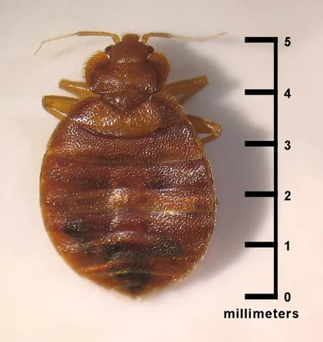
Adult bed bug beside a millimeter scale. Photo: CDC / CDC-DPDx; Blaine Mathison. Public domain (U.S. government work). Source: CDC PHIL; original image available free from CDC. Use does not imply CDC, HHS, or U.S. Government endorsement.
Adult bed bug beside a millimeter scale.
Adult specimen only. Nymphs and recently fed bugs look different. The printed scale describes this specimen, not the image size on a screen. Bites alone cannot establish bed bugs.
Original source (new tab) · Public domain (U.S. government work)
Changes: Resized and encoded as WebP; composition and substantive visual content preserved. Derivative image licence: Public domain (U.S. government work).
German cockroach, archival dorsal photograph
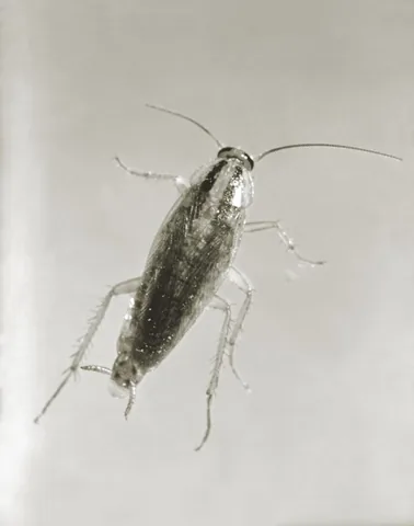
German cockroach, archival dorsal photograph. Photo: CDC. Public domain (U.S. government work). Source: CDC PHIL; original image available free from CDC. Use does not imply CDC, HHS, or U.S. Government endorsement.
German cockroach, archival dorsal photograph.
Monochrome archival view. Use the color image for coloration. A dorsal photograph alone cannot reliably separate German and Asian cockroaches.
Original source (new tab) · Public domain (U.S. government work)
Changes: Resized and encoded as WebP; composition and substantive visual content preserved. Derivative image licence: Public domain (U.S. government work).
Adult female blacklegged tick on a leaf
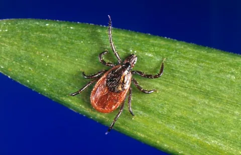
Adult female blacklegged tick on a leaf. Photo: Jim Gathany; CDC / Michael L. Levin. Public domain (U.S. government work). Source: CDC PHIL; original image available free from CDC. Use does not imply CDC, HHS, or U.S. Government endorsement.
Adult female blacklegged tick on a leaf.
One adult female; not a plate for all tick species or stages. Engorgement changes appearance. Do not infer infection or exposure risk from the image.
Original source (new tab) · Public domain (U.S. government work)
Changes: Resized and encoded as WebP; composition and substantive visual content preserved. Derivative image licence: Public domain (U.S. government work).
Adult Indianmeal moth on cereal material
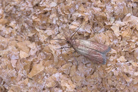
Adult Indianmeal moth on cereal material. Photo: Scott Bauer, USDA Agricultural Research Service. Public domain (U.S. government work). Use does not imply USDA ARS endorsement.
Adult Indianmeal moth on cereal material.
Adult only. The background is illustrative of stored food and does not establish which food in a home is infested.
Original source (new tab) · Public domain (U.S. government work)
Changes: Resized and encoded as WebP; composition and substantive visual content preserved. Derivative image licence: Public domain (U.S. government work).
Formosan termite swarmers captured on a monitoring trap
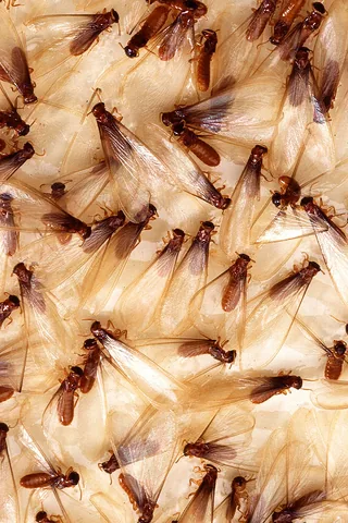
Formosan termite swarmers captured on a monitoring trap. Photo: Scott Bauer, USDA Agricultural Research Service. Public domain (U.S. government work). Use does not imply USDA ARS endorsement.
Formosan termite swarmers captured on a monitoring trap.
Multiple trapped swarmers with overlapping wings. This is a Formosan termite example, not a diagnostic plate for all termites; a swarmer does not establish the extent of structural infestation.
Original source (new tab) · Public domain (U.S. government work)
Changes: Resized and encoded as WebP; composition and substantive visual content preserved. Derivative image licence: Public domain (U.S. government work).
Odorous house ant
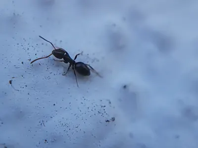
Odorous house ant. Photo: Theo Summer. CC BY 4.0.
Odorous house ant.
One small ant species, not a plate for all household ants. Fine waist characters are not resolved; confirm size, behavior, location and a regional key before choosing a treatment.
Original source (new tab) · CC BY 4.0
Changes: Resized and encoded as WebP; full source composition and substantive visual content preserved. No crop, recoloring, retouching or generated diagnostic content added. Derivative image licence: CC BY 4.0.
Eastern black carpenter ant
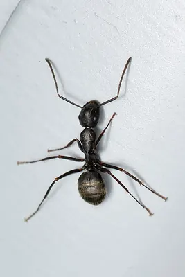
Eastern black carpenter ant. Photo: Ryan Hodnett. CC BY-SA 4.0.
Eastern black carpenter ant.
One North American carpenter ant example. Other carpenter ants differ in color and size. The image does not establish nesting in a building or distinguish every similar ant.
Original source (new tab) · CC BY-SA 4.0
Changes: Resized and encoded as WebP; full source composition and substantive visual content preserved. No crop, recoloring, retouching or generated diagnostic content added. Downloaded a Wikimedia-generated display-size copy because the original CDN requested standard thumbnail sizes; downloaded-source dimensions and hash are recorded separately. Derivative image licence: CC BY-SA 4.0.
Red imported fire ant worker foraging
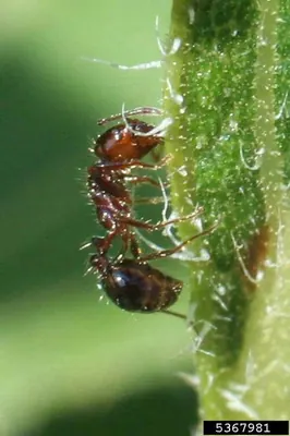
Red imported fire ant worker foraging. Photo: Russ Ottens / Forestry Images. CC BY 3.0 us.
Red imported fire ant worker foraging.
One worker example. Other stinging ants and imported-fire-ant hybrids can look similar. Do not handle ants or disturb a mound to reproduce this view.
Original source (new tab) · CC BY 3.0 us
Changes: Resized and encoded as WebP; full source composition and substantive visual content preserved. No crop, recoloring, retouching or generated diagnostic content added. Downloaded a Wikimedia-generated display-size copy because the original CDN requested standard thumbnail sizes; downloaded-source dimensions and hash are recorded separately. Derivative image licence: CC BY 3.0 us.
Brown powderpost beetle, dorsal specimen view
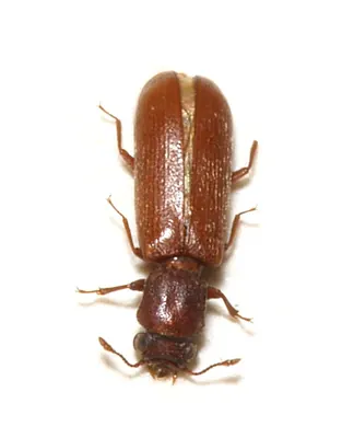
Brown powderpost beetle, dorsal specimen view. Photo: Sarefo. CC BY-SA 3.0.
Brown powderpost beetle, dorsal specimen view.
A magnified adult of one lyctine species, not a key for every wood borer. Adult identity, wood type, hole and frass evidence, and current activity must agree.
Original source (new tab) · CC BY-SA 3.0
Changes: Resized and encoded as WebP; full source composition and substantive visual content preserved. No crop, recoloring, retouching or generated diagnostic content added. Derivative image licence: CC BY-SA 3.0.
Common furniture beetle, dorsal specimen view
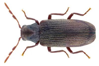
Common furniture beetle, dorsal specimen view. Photo: Udo Schmidt from Deutschland. CC BY-SA 2.0.
Common furniture beetle, dorsal specimen view.
One preserved adult. Similar wood-boring beetles require fine characters; an adult photograph cannot date exit holes or establish active structural damage.
Original source (new tab) · CC BY-SA 2.0
Changes: Resized and encoded as WebP; full source composition and substantive visual content preserved. No crop, recoloring, retouching or generated diagnostic content added. Downloaded a Wikimedia-generated display-size copy because the original CDN requested standard thumbnail sizes; downloaded-source dimensions and hash are recorded separately. Derivative image licence: CC BY-SA 2.0.
Female eastern carpenter bee, side view
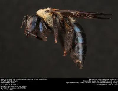
Female eastern carpenter bee, side view. Photo: Alejandro Santillana / Insects Unlocked, University of Texas at Austin. CC0.
Female eastern carpenter bee, side view.
A female example with a relatively shiny abdomen. Other carpenter bees vary, and the photograph does not show a nest entrance or prove damage to a particular timber.
Original source (new tab) · CC0
Changes: Resized and encoded as WebP; full source composition and substantive visual content preserved. No crop, recoloring, retouching or generated diagnostic content added. Derivative image licence: CC0.
Two buff-tailed bumblebees on flowers
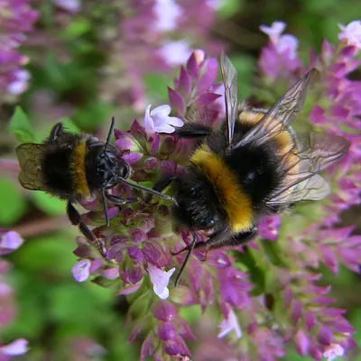
Two buff-tailed bumblebees on flowers. Photo: linsepatron. CC BY 2.0.
Two buff-tailed bumblebees on flowers.
Source labels a small female worker at left and larger male at right. This European bumblebee example illustrates a hairy abdomen; it is not a key to North American bumblebees or every bee species.
Original source (new tab) · CC BY 2.0
Changes: Resized and encoded as WebP; full source composition and substantive visual content preserved. No crop, recoloring, retouching or generated diagnostic content added. Downloaded a Wikimedia-generated display-size copy because the original CDN requested standard thumbnail sizes; downloaded-source dimensions and hash are recorded separately. Derivative image licence: CC BY 2.0.
Webbing clothes moth at rest
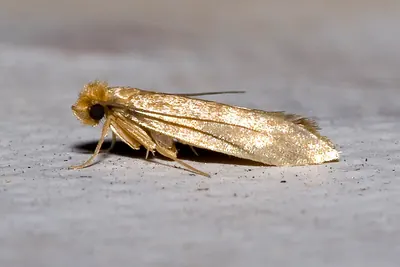
Webbing clothes moth at rest. Photo: Olaf Leillinger. CC BY-SA 2.5.
Webbing clothes moth at rest.
One adult moth with folded wings. Confirm larval activity in susceptible fabric; an adult alone does not prove fabric damage, and lighting affects apparent buff coloration.
Original source (new tab) · CC BY-SA 2.5
Changes: Resized and encoded as WebP; full source composition and substantive visual content preserved. No crop, recoloring, retouching or generated diagnostic content added. Downloaded a Wikimedia-generated display-size copy because the original CDN requested standard thumbnail sizes; downloaded-source dimensions and hash are recorded separately. Derivative image licence: CC BY-SA 2.5.
Case-bearing clothes moth at rest
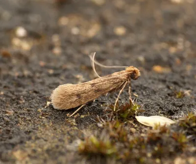
Case-bearing clothes moth at rest. Photo: Ben Sale from UK. CC BY 2.0.
Case-bearing clothes moth at rest.
One adult of a second clothes-moth species. Adults can be confused with other small moths; inspect fabric and seek the portable larval case rather than relying on color alone.
Original source (new tab) · CC BY 2.0
Changes: Resized and encoded as WebP; full source composition and substantive visual content preserved. No crop, recoloring, retouching or generated diagnostic content added. Downloaded a Wikimedia-generated display-size copy because the original CDN requested standard thumbnail sizes; downloaded-source dimensions and hash are recorded separately. Derivative image licence: CC BY 2.0.
Case-bearing clothes moth larva with its case
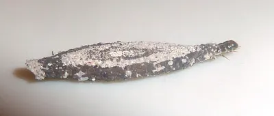
Case-bearing clothes moth larva with its case. Photo: JMK. CC BY-SA 4.0.
Case-bearing clothes moth larva with its case.
A larva photographed in Pretoria, South Africa. Fibers affect the case color; other case-bearing insects differ. A photograph alone does not establish the source of fabric damage.
Original source (new tab) · CC BY-SA 4.0
Changes: Resized and encoded as WebP; full source composition and substantive visual content preserved. No crop, recoloring, retouching or generated diagnostic content added. Downloaded a Wikimedia-generated display-size copy because the original CDN requested standard thumbnail sizes; downloaded-source dimensions and hash are recorded separately. Derivative image licence: CC BY-SA 4.0.
Adult varied carpet beetle on a flower
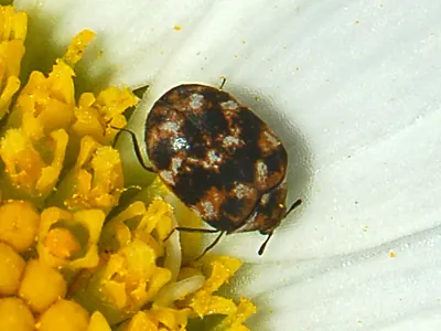
Adult varied carpet beetle on a flower. Photo: Hectonichus. CC BY-SA 3.0.
Adult varied carpet beetle on a flower.
An adult of one carpet beetle species. Adults often occur at flowers or windows; inspect for larvae and a food source before concluding that fabric is infested.
Original source (new tab) · CC BY-SA 3.0
Changes: Resized and encoded as WebP; full source composition and substantive visual content preserved. No crop, recoloring, retouching or generated diagnostic content added. Derivative image licence: CC BY-SA 3.0.
Varied carpet beetle larva, side view
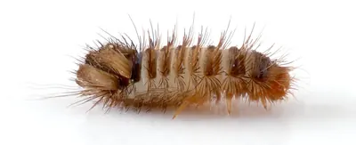
Varied carpet beetle larva, side view. Photo: André Karwath aka Aka. CC BY-SA 2.5.
Varied carpet beetle larva, side view.
Magnified hairy larva. Other dermestid larvae vary, and this specimen does not identify a skin reaction. Bed bugs are flattened insects with a different outline; retain a specimen when uncertain.
Original source (new tab) · CC BY-SA 2.5
Changes: Resized and encoded as WebP; full source composition and substantive visual content preserved. No crop, recoloring, retouching or generated diagnostic content added. Derivative image licence: CC BY-SA 2.5.
Common silverfish on a wall
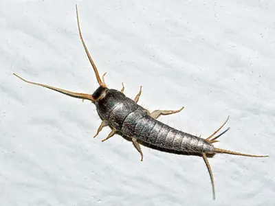
Common silverfish on a wall. Photo: Christian Fischer. CC BY-SA 3.0.
Common silverfish on a wall.
A common silverfish example with three tail filaments. Body size on a screen is meaningless without a scale; other silverfish and young stages need separate identification.
Original source (new tab) · CC BY-SA 3.0
Changes: Resized and encoded as WebP; full source composition and substantive visual content preserved. No crop, recoloring, retouching or generated diagnostic content added. Downloaded a Wikimedia-generated display-size copy because the original CDN requested standard thumbnail sizes; downloaded-source dimensions and hash are recorded separately. Derivative image licence: CC BY-SA 3.0.
Firebrat photographed under a microscope
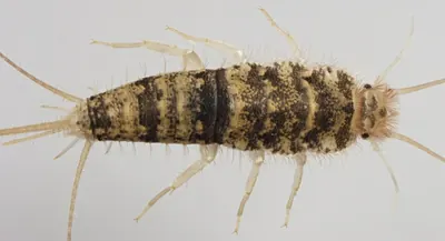
Firebrat photographed under a microscope. Photo: KyleCMSmith. CC BY-SA 4.0.
Firebrat photographed under a microscope.
Source specimen was found in a North Carolina boiler room. Mottled coloration is an example, not a substitute for confirming warm habitat and the insect's structure.
Original source (new tab) · CC BY-SA 4.0
Changes: Resized and encoded as WebP; full source composition and substantive visual content preserved. No crop, recoloring, retouching or generated diagnostic content added. Downloaded a Wikimedia-generated display-size copy because the original CDN requested standard thumbnail sizes; downloaded-source dimensions and hash are recorded separately. Derivative image licence: CC BY-SA 4.0.
Long-tailed silverfish on a dark background
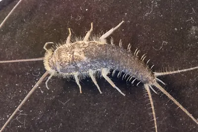
Long-tailed silverfish on a dark background. Photo: Rene Sylvestersen. CC BY-SA 4.0.
Long-tailed silverfish on a dark background.
One long-tailed silverfish example. Some tail filaments extend beyond this source frame; do not measure tail-to-body ratios from it. Tail filaments and antennae are delicate on household specimens; color alone cannot separate silverfish species.
Original source (new tab) · CC BY-SA 4.0
Changes: Resized and encoded as WebP; full source composition and substantive visual content preserved. No crop, recoloring, retouching or generated diagnostic content added. Downloaded a Wikimedia-generated display-size copy because the original CDN requested standard thumbnail sizes; downloaded-source dimensions and hash are recorded separately. Derivative image licence: CC BY-SA 4.0.
Adult booklouse under magnification
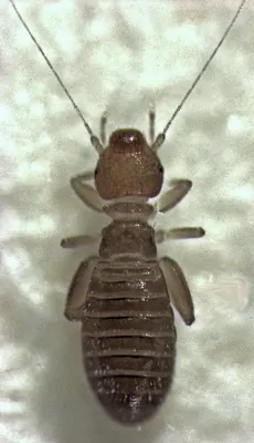
Adult booklouse under magnification. Photo: S.E. Thorpe. Public domain.
Adult booklouse under magnification.
Genus-level specimen photographed indoors in New Zealand. It is not identified as a particular Liposcelis species; no screen image establishes its real size or the source of a moisture problem.
Original source (new tab) · Public domain

Changes: Resized and encoded as WebP; full source composition and substantive visual content preserved. No crop, recoloring, retouching or generated diagnostic content added. Derivative image licence: Public domain.
Drain fly with hairy wings
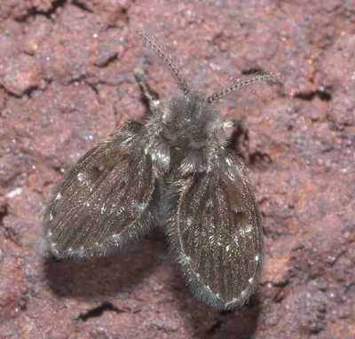
Drain fly with hairy wings. Photo: xpda. CC BY-SA 4.0.
Drain fly with hairy wings.
One moth-fly species. The resting shape and hairy wings help distinguish this group, but the photo cannot identify which drain or other wet organic source is producing flies.
Original source (new tab) · CC BY-SA 4.0
Changes: Resized and encoded as WebP; full source composition and substantive visual content preserved. No crop, recoloring, retouching or generated diagnostic content added. Downloaded a Wikimedia-generated display-size copy because the original CDN requested standard thumbnail sizes; downloaded-source dimensions and hash are recorded separately. Derivative image licence: CC BY-SA 4.0.
Fruit fly, dorsal view
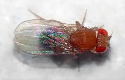
Fruit fly, dorsal view. Photo: André Karwath aka Aka. CC BY-SA 2.5.
Fruit fly, dorsal view.
A magnified adult fruit fly. Not all small flies have red eyes. Confirm the body shape and breeding material; this does not identify every vinegar fly or establish a source.
Original source (new tab) · CC BY-SA 2.5
Changes: Resized and encoded as WebP; full source composition and substantive visual content preserved. No crop, recoloring, retouching or generated diagnostic content added. Derivative image licence: CC BY-SA 2.5.
Fungus gnat, side view
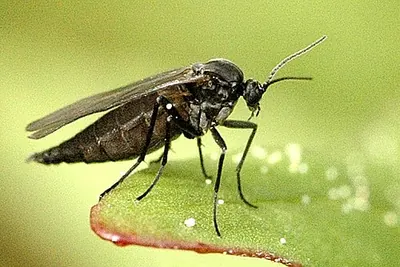
Fungus gnat, side view. Photo: James K. Lindsey. CC BY-SA 2.5.
Fungus gnat, side view.
One fungus gnat species photographed in Belgium. Long legs and a slender body are visible, but species-level identification needs finer characters and the breeding source must be checked separately.
Original source (new tab) · CC BY-SA 2.5
Changes: Resized and encoded as WebP; full source composition and substantive visual content preserved. No crop, recoloring, retouching or generated diagnostic content added. Downloaded a Wikimedia-generated display-size copy because the original CDN requested standard thumbnail sizes; downloaded-source dimensions and hash are recorded separately. Derivative image licence: CC BY-SA 2.5.
Common house fly
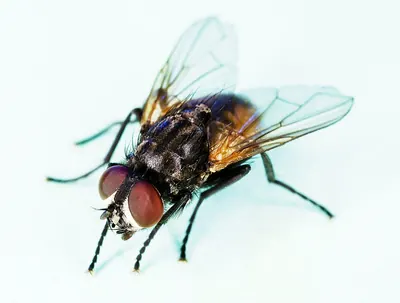
Common house fly. Photo: USDAgov. CC BY 2.0.
Common house fly.
One adult house fly. Similar flies and biting flies need separate identification; this image does not prove a sanitation source or show every character used in a fly key.
Original source (new tab) · CC BY 2.0
Changes: Resized and encoded as WebP; full source composition and substantive visual content preserved. No crop, recoloring, retouching or generated diagnostic content added. Downloaded a Wikimedia-generated display-size copy because the original CDN requested standard thumbnail sizes; downloaded-source dimensions and hash are recorded separately. Derivative image licence: CC BY 2.0.
Cluster fly on a flower
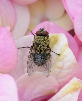
Cluster fly on a flower. Photo: Alvesgaspar. CC BY-SA 3.0.
Cluster fly on a flower.
An adult photographed outside. Golden hairs can be worn or hard to see on a household specimen; an indoor fly is not automatically a cluster fly or evidence of indoor breeding.
Original source (new tab) · CC BY-SA 3.0
Changes: Resized and encoded as WebP; full source composition and substantive visual content preserved. No crop, recoloring, retouching or generated diagnostic content added. Downloaded a Wikimedia-generated display-size copy because the original CDN requested standard thumbnail sizes; downloaded-source dimensions and hash are recorded separately. Derivative image licence: CC BY-SA 3.0.
Yellow fever mosquito, Aedes aegypti
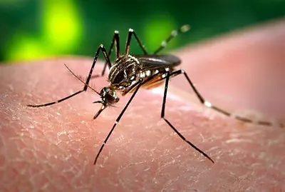
Yellow fever mosquito, Aedes aegypti. Photo: James Gathany. Public domain. Source: CDC PHIL; original image available free from CDC. Use does not imply CDC, HHS, or U.S. Government endorsement.
Yellow fever mosquito, Aedes aegypti.
One mosquito species and sex, not a complete regional mosquito key. Other mosquitoes look different. An image cannot determine whether a mosquito carries a pathogen or whether exposure occurred.
Original source (new tab) · Public domain
Changes: Resized and encoded as WebP; full source composition and substantive visual content preserved. No crop, recoloring, retouching or generated diagnostic content added. Downloaded a Wikimedia-generated display-size copy because the original CDN requested standard thumbnail sizes; downloaded-source dimensions and hash are recorded separately. Derivative image licence: Public domain.
Male human head louse on hair
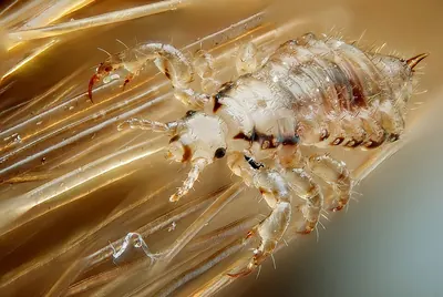
Male human head louse on hair. Photo: Gilles San Martin. CC BY-SA 2.0.
Male human head louse on hair.
Magnified male head louse. Body lice and pubic lice require a different assessment; a nit, skin mark or sensation alone does not confirm live head lice.
Original source (new tab) · CC BY-SA 2.0
Changes: Resized and encoded as WebP; full source composition and substantive visual content preserved. No crop, recoloring, retouching or generated diagnostic content added. Downloaded a Wikimedia-generated display-size copy because the original CDN requested standard thumbnail sizes; downloaded-source dimensions and hash are recorded separately. Derivative image licence: CC BY-SA 2.0.
Clover mite under a digital microscope
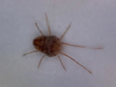
Clover mite under a digital microscope. Photo: PerAsperaAdInferos. CC BY-SA 4.0.
Clover mite under a digital microscope.
A greatly magnified clover mite example. This plant-feeding mite must not be relabeled as a bird mite, rodent mite, scabies mite or cause of bites. Color alone does not identify a mite.
Original source (new tab) · CC BY-SA 4.0
Changes: Resized and encoded as WebP; full source composition and substantive visual content preserved. No crop, recoloring, retouching or generated diagnostic content added. Downloaded a Wikimedia-generated display-size copy because the original CDN requested standard thumbnail sizes; downloaded-source dimensions and hash are recorded separately. Derivative image licence: CC BY-SA 4.0.
Poultry red mite under magnification
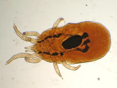
Poultry red mite under magnification. Photo: Luis Fernández García. CC BY-SA 2.5 es.
Poultry red mite under magnification.
A photomicrograph of one bird-associated mite. Fine microscopic characters and a source investigation are needed for identification; it cannot diagnose human skin symptoms or represent all mites.
Original source (new tab) · CC BY-SA 2.5 es
Changes: Resized and encoded as WebP; full source composition and substantive visual content preserved. No crop, recoloring, retouching or generated diagnostic content added. Downloaded a Wikimedia-generated display-size copy because the original CDN requested standard thumbnail sizes; downloaded-source dimensions and hash are recorded separately. Derivative image licence: CC BY-SA 2.5 es.
European paper wasp
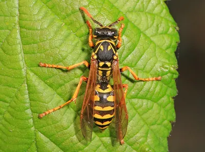
European paper wasp. Photo: Judy Gallagher. CC BY 2.0.
European paper wasp.
One paper-wasp species. Nest type and location matter, and other paper wasps vary in color. Do not approach or disturb a nest to match this photograph.
Original source (new tab) · CC BY 2.0
Changes: Resized and encoded as WebP; full source composition and substantive visual content preserved. No crop, recoloring, retouching or generated diagnostic content added. Downloaded a Wikimedia-generated display-size copy because the original CDN requested standard thumbnail sizes; downloaded-source dimensions and hash are recorded separately. Derivative image licence: CC BY 2.0.
German yellowjacket on a flower
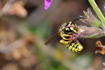
German yellowjacket on a flower. Photo: Manuel Hervas. CC BY-SA 4.0.
German yellowjacket on a flower.
One yellowjacket species. Markings vary and photographs do not reliably identify every yellowjacket; follow the nest and flight-path evidence from a safe distance.
Original source (new tab) · CC BY-SA 4.0
Changes: Resized and encoded as WebP; full source composition and substantive visual content preserved. No crop, recoloring, retouching or generated diagnostic content added. Downloaded a Wikimedia-generated display-size copy because the original CDN requested standard thumbnail sizes; downloaded-source dimensions and hash are recorded separately. Derivative image licence: CC BY-SA 4.0.
Western honey bee on a flower
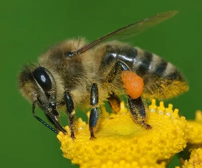
Western honey bee on a flower. Photo: Andreas Trepte. CC BY-SA 2.5.
Western honey bee on a flower.
One honey bee worker. It does not represent bumblebees or solitary bees, and it cannot establish whether an observed cluster is a temporary swarm or an established colony.
Original source (new tab) · CC BY-SA 2.5
Changes: Resized and encoded as WebP; full source composition and substantive visual content preserved. No crop, recoloring, retouching or generated diagnostic content added. Downloaded a Wikimedia-generated display-size copy because the original CDN requested standard thumbnail sizes; downloaded-source dimensions and hash are recorded separately. Derivative image licence: CC BY-SA 2.5.
Female southern black widow, underside view
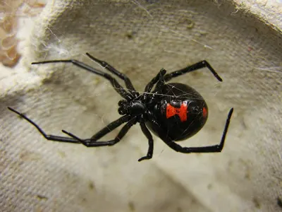
Female southern black widow, underside view. Photo: Shenrich91. CC BY-SA 3.0.
Female southern black widow, underside view.
Source labels an adult gravid female. The red hourglass is shown on the underside, which may not be safely visible on a live spider. Other widow species and immature stages differ; do not handle one for identification.
Original source (new tab) · CC BY-SA 3.0
Changes: Resized and encoded as WebP; full source composition and substantive visual content preserved. No crop, recoloring, retouching or generated diagnostic content added. Downloaded a Wikimedia-generated display-size copy because the original CDN requested standard thumbnail sizes; downloaded-source dimensions and hash are recorded separately. Derivative image licence: CC BY-SA 3.0.
Brown recluse spider
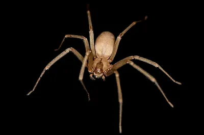
Brown recluse spider. Photo: Rosa Pineda. CC BY-SA 3.0.
Brown recluse spider.
One recluse spider example. A violin-like mark alone is unreliable, and six-eye arrangement is not a practical household photograph test. Range, a specimen and expert identification matter; no spider image diagnoses a wound.
Original source (new tab) · CC BY-SA 3.0
Changes: Resized and encoded as WebP; full source composition and substantive visual content preserved. No crop, recoloring, retouching or generated diagnostic content added. Downloaded a Wikimedia-generated display-size copy because the original CDN requested standard thumbnail sizes; downloaded-source dimensions and hash are recorded separately. Derivative image licence: CC BY-SA 3.0.
Common house spider
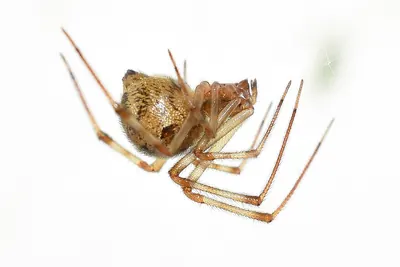
Common house spider. Photo: Daniel.fox123. CC BY 4.0.
Common house spider.
One common cobweb spider example. Females, males and young differ; many harmless spiders resemble one another. This photo does not identify a skin reaction or establish that a spider caused it.
Original source (new tab) · CC BY 4.0
Changes: Resized and encoded as WebP; full source composition and substantive visual content preserved. No crop, recoloring, retouching or generated diagnostic content added. Downloaded a Wikimedia-generated display-size copy because the original CDN requested standard thumbnail sizes; downloaded-source dimensions and hash are recorded separately. Derivative image licence: CC BY 4.0.
Arizona bark scorpion
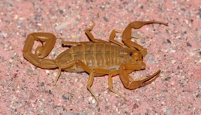
Arizona bark scorpion. Photo: Andrew Meeds. CC BY 4.0.
Arizona bark scorpion.
One bark-scorpion example. A single photograph cannot reliably separate all scorpions or predict sting severity. Do not handle a scorpion; seek regional identification and prompt sting advice when relevant.
Original source (new tab) · CC BY 4.0
Changes: Resized and encoded as WebP; full source composition and substantive visual content preserved. No crop, recoloring, retouching or generated diagnostic content added. Downloaded a Wikimedia-generated display-size copy because the original CDN requested standard thumbnail sizes; downloaded-source dimensions and hash are recorded separately. Derivative image licence: CC BY 4.0.
Eastern bloodsucking conenose, dorsal view
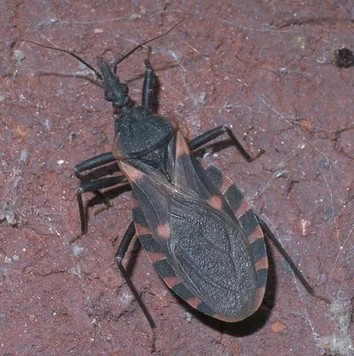
Eastern bloodsucking conenose, dorsal view. Photo: xpda. CC BY-SA 4.0.
Eastern bloodsucking conenose, dorsal view.
One North American triatomine species. Other kissing bugs differ. Fine head and body characters, location and specimen confirmation matter; the image cannot establish infection or transmission.
Original source (new tab) · CC BY-SA 4.0
Changes: Resized and encoded as WebP; full source composition and substantive visual content preserved. No crop, recoloring, retouching or generated diagnostic content added. Downloaded a Wikimedia-generated display-size copy because the original CDN requested standard thumbnail sizes; downloaded-source dimensions and hash are recorded separately. Derivative image licence: CC BY-SA 4.0.
Adult wheel bug, a kissing-bug lookalike
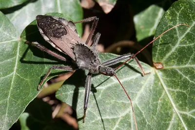
Adult wheel bug, a kissing-bug lookalike. Photo: Ryan Kaldari. Public domain.
Adult wheel bug, a kissing-bug lookalike.
Adult wheel bug showing its raised coglike crest. Wheel-bug nymphs lack this adult crest. This predatory assassin bug is a lookalike, not a triatomine; do not handle either group.
Original source (new tab) · Public domain

Changes: Resized and encoded as WebP; full source composition and substantive visual content preserved. No crop, recoloring, retouching or generated diagnostic content added. Downloaded a Wikimedia-generated display-size copy because the original CDN requested standard thumbnail sizes; downloaded-source dimensions and hash are recorded separately. Derivative image licence: Public domain.
House centipede, dorsal view
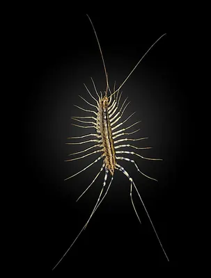
House centipede, dorsal view. Photo: Didier Descouens. CC BY-SA 4.0.
House centipede, dorsal view.
One living house centipede with long legs. It is not a millipede or woodlouse. The image does not establish how many are present or identify the underlying moisture and prey sources.
Original source (new tab) · CC BY-SA 4.0
Changes: Resized and encoded as WebP; full source composition and substantive visual content preserved. No crop, recoloring, retouching or generated diagnostic content added. Downloaded a Wikimedia-generated display-size copy because the original CDN requested standard thumbnail sizes; downloaded-source dimensions and hash are recorded separately. Derivative image licence: CC BY-SA 4.0.
Rough woodlouse, side view
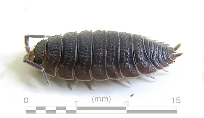
Rough woodlouse, side view. Photo: Yug. CC BY-SA 3.0.
Rough woodlouse, side view.
An adult terrestrial crustacean of one species. Pillbugs and sowbugs differ, and this image does not show every species or prove a dampness defect indoors.
Original source (new tab) · CC BY-SA 3.0
Changes: Resized and encoded as WebP; full source composition and substantive visual content preserved. No crop, recoloring, retouching or generated diagnostic content added. Downloaded a Wikimedia-generated display-size copy because the original CDN requested standard thumbnail sizes; downloaded-source dimensions and hash are recorded separately. Derivative image licence: CC BY-SA 3.0.
Greenhouse millipede
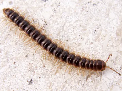
Greenhouse millipede. Photo: JCoelho. CC BY 2.0.
Greenhouse millipede.
One millipede species photographed in Portugal. The many-legged cylindrical form differs from a centipede; species identification and the source of an indoor influx need separate checks.
Original source (new tab) · CC BY 2.0
Changes: Resized and encoded as WebP; full source composition and substantive visual content preserved. No crop, recoloring, retouching or generated diagnostic content added. Downloaded a Wikimedia-generated display-size copy because the original CDN requested standard thumbnail sizes; downloaded-source dimensions and hash are recorded separately. Derivative image licence: CC BY 2.0.
Springtail, Orchesella cincta
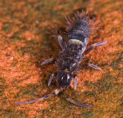
Springtail, Orchesella cincta. Photo: Mvuijlst. CC BY-SA 3.0.
Springtail, Orchesella cincta.
A visibly marked springtail species photographed in Belgium. Many household springtails are paler or rounder. Jumping behavior and magnified structure help; the image alone does not identify a moisture source.
Original source (new tab) · CC BY-SA 3.0
Changes: Resized and encoded as WebP; full source composition and substantive visual content preserved. No crop, recoloring, retouching or generated diagnostic content added. Downloaded a Wikimedia-generated display-size copy because the original CDN requested standard thumbnail sizes; downloaded-source dimensions and hash are recorded separately. Derivative image licence: CC BY-SA 3.0.
European earwig
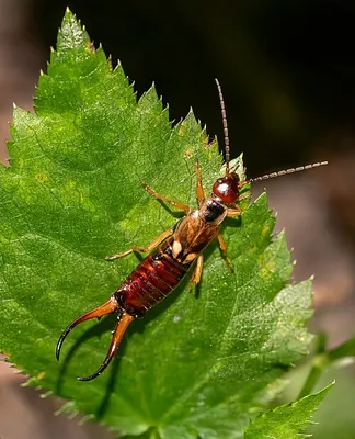
European earwig. Photo: Rhododendrites. CC BY-SA 4.0.
European earwig.
One adult earwig showing rear forceps. Forceps shape varies by sex and stage; the presence of an earwig does not establish that it caused observed plant or fabric damage.
Original source (new tab) · CC BY-SA 4.0
Changes: Resized and encoded as WebP; full source composition and substantive visual content preserved. No crop, recoloring, retouching or generated diagnostic content added. Downloaded a Wikimedia-generated display-size copy because the original CDN requested standard thumbnail sizes; downloaded-source dimensions and hash are recorded separately. Derivative image licence: CC BY-SA 4.0.
Adult female house cricket
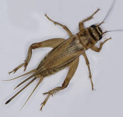
Adult female house cricket. Photo: Geyersberg, Professor emeritus Hans Schneider. CC BY-SA 3.0.
Adult female house cricket.
An adult female with a central egg-laying ovipositor between the rear cerci. The ovipositor is not a stinger. Other crickets and immature stages differ.
Original source (new tab) · CC BY-SA 3.0
Changes: Resized and encoded as WebP; full source composition and substantive visual content preserved. No crop, recoloring, retouching or generated diagnostic content added. Downloaded a Wikimedia-generated display-size copy because the original CDN requested standard thumbnail sizes; downloaded-source dimensions and hash are recorded separately. Derivative image licence: CC BY-SA 3.0.
Immature greenhouse camel cricket, side view
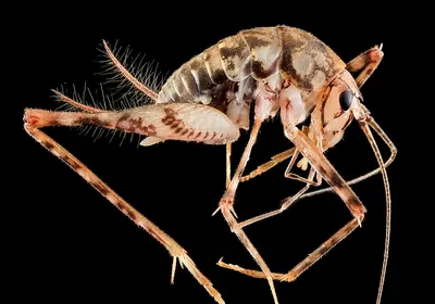
Immature greenhouse camel cricket, side view. Photo: USGS Bee Inventory and Monitoring Lab. CC BY 2.0.
Immature greenhouse camel cricket, side view.
Source labels an immature camel cricket from a Maryland basement. Adult size and coloration differ; it is an insect with six legs, not a spider, and it has no true wings.
Original source (new tab) · CC BY 2.0
Changes: Resized and encoded as WebP; full source composition and substantive visual content preserved. No crop, recoloring, retouching or generated diagnostic content added. Downloaded a Wikimedia-generated display-size copy because the original CDN requested standard thumbnail sizes; downloaded-source dimensions and hash are recorded separately. Derivative image licence: CC BY 2.0.
Adult brown marmorated stink bug
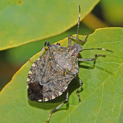
Adult brown marmorated stink bug. Photo: Hectonichus. CC BY-SA 4.0.
Adult brown marmorated stink bug.
One adult. Other shield-shaped bugs can resemble it; check antenna and edge markings with a regional key. Its presence indoors does not establish indoor breeding.
Original source (new tab) · CC BY-SA 4.0
Changes: Resized and encoded as WebP; full source composition and substantive visual content preserved. No crop, recoloring, retouching or generated diagnostic content added. Downloaded a Wikimedia-generated display-size copy because the original CDN requested standard thumbnail sizes; downloaded-source dimensions and hash are recorded separately. Derivative image licence: CC BY-SA 4.0.
Adult eastern boxelder bug
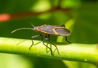
Adult eastern boxelder bug. Photo: Rhododendrites. CC BY-SA 4.0.
Adult eastern boxelder bug.
One adult photographed in New York. Nymphs are smaller, redder and lack full wings. Other red-and-black bugs require different identification; this image is not a key for all nuisance bugs.
Original source (new tab) · CC BY-SA 4.0
Changes: Resized and encoded as WebP; full source composition and substantive visual content preserved. No crop, recoloring, retouching or generated diagnostic content added. Downloaded a Wikimedia-generated display-size copy because the original CDN requested standard thumbnail sizes; downloaded-source dimensions and hash are recorded separately. Derivative image licence: CC BY-SA 4.0.
Multicolored Asian lady beetle, dorsal specimen view
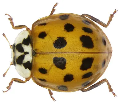
Multicolored Asian lady beetle, dorsal specimen view. Photo: URSchmidt. CC BY-SA 4.0.
Multicolored Asian lady beetle, dorsal specimen view.
A preserved adult showing one color form. Spot number and color vary widely, and other lady beetles can resemble it. Do not identify this species from orange color alone.
Original source (new tab) · CC BY-SA 4.0
Changes: Resized and encoded as WebP; full source composition and substantive visual content preserved. No crop, recoloring, retouching or generated diagnostic content added. Downloaded a Wikimedia-generated display-size copy because the original CDN requested standard thumbnail sizes; downloaded-source dimensions and hash are recorded separately. Derivative image licence: CC BY-SA 4.0.
Male green drake mayfly
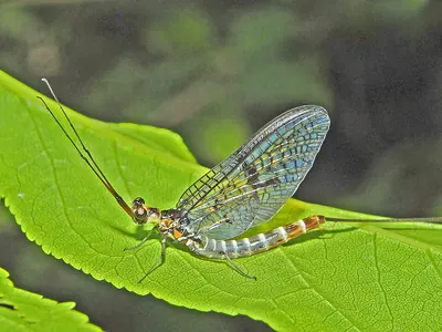
Male green drake mayfly. Photo: Hectonichus. CC BY-SA 3.0.
Male green drake mayfly.
A male mayfly photographed in Italy. Tail filaments extend beyond this source frame, so tail count is not shown completely. Other mayflies vary in wing shape and tail count; this is an example of seasonal aquatic insects, not a key to all emerging flies or evidence of indoor breeding.
Original source (new tab) · CC BY-SA 3.0
Changes: Resized and encoded as WebP; full source composition and substantive visual content preserved. No crop, recoloring, retouching or generated diagnostic content added. Downloaded a Wikimedia-generated display-size copy because the original CDN requested standard thumbnail sizes; downloaded-source dimensions and hash are recorded separately. Derivative image licence: CC BY-SA 3.0.
Periodical cicadas
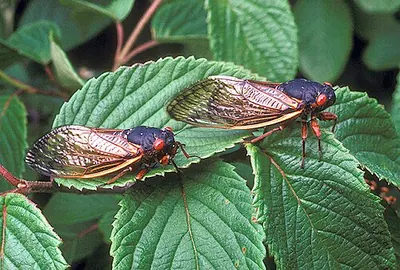
Periodical cicadas. Photo: ARS Information Staff / USDA Agricultural Research Service. Public domain. Use does not imply USDA ARS endorsement.
Periodical cicadas.
One eastern North American periodical cicada species. Local brood timing, annual cicadas and other species differ; the image does not predict a local emergence year.
Original source (new tab) · Public domain

Changes: Resized and encoded as WebP; full source composition and substantive visual content preserved. No crop, recoloring, retouching or generated diagnostic content added. Downloaded a Wikimedia-generated display-size copy because the original CDN requested standard thumbnail sizes; downloaded-source dimensions and hash are recorded separately. Derivative image licence: Public domain.
Male nonbiting midge
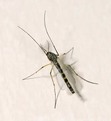
Male nonbiting midge. Photo: Didier Descouens. CC BY-SA 4.0.
Male nonbiting midge.
A male with feathery antennae. Other midge species and females differ. This example helps distinguish a seasonal midge from mosquitoes, but no single image establishes the identity of every small swarming fly.
Original source (new tab) · CC BY-SA 4.0
Changes: Resized and encoded as WebP; full source composition and substantive visual content preserved. No crop, recoloring, retouching or generated diagnostic content added. Downloaded a Wikimedia-generated display-size copy because the original CDN requested standard thumbnail sizes; downloaded-source dimensions and hash are recorded separately. Derivative image licence: CC BY-SA 4.0.
Drugstore beetle, dorsal specimen view
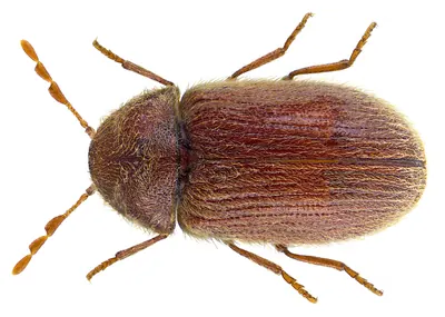
Drugstore beetle, dorsal specimen view. Photo: Udo Schmidt from Deutschland. CC BY-SA 2.0.
Drugstore beetle, dorsal specimen view.
One magnified stored-product beetle. The longitudinal wing-cover rows and clubbed antennae are fine characters; inspect suspect products and distinguish cigarette beetles or furniture beetles before choosing action.
Original source (new tab) · CC BY-SA 2.0
Changes: Resized and encoded as WebP; full source composition and substantive visual content preserved. No crop, recoloring, retouching or generated diagnostic content added. Downloaded a Wikimedia-generated display-size copy because the original CDN requested standard thumbnail sizes; downloaded-source dimensions and hash are recorded separately. Derivative image licence: CC BY-SA 2.0.
Cigarette beetle, dorsal specimen view
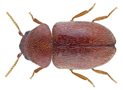
Cigarette beetle, dorsal specimen view. Photo: Udo Schmidt from Deutschland. CC BY-SA 2.0.
Cigarette beetle, dorsal specimen view.
One magnified adult. Fine serrated antennae and wing-cover texture help separate it from the drugstore beetle; overall brown color and a rounded outline alone are insufficient.
Original source (new tab) · CC BY-SA 2.0
Changes: Resized and encoded as WebP; full source composition and substantive visual content preserved. No crop, recoloring, retouching or generated diagnostic content added. Downloaded a Wikimedia-generated display-size copy because the original CDN requested standard thumbnail sizes; downloaded-source dimensions and hash are recorded separately. Derivative image licence: CC BY-SA 2.0.
Rice weevil, dorsal specimen view
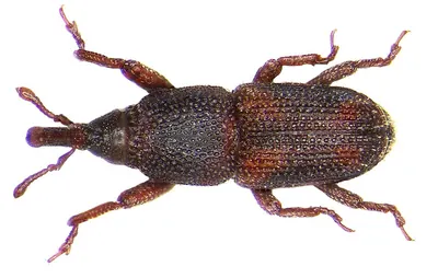
Rice weevil, dorsal specimen view. Photo: Udo Schmidt from Deutschland. CC BY-SA 2.0.
Rice weevil, dorsal specimen view.
One preserved snouted grain weevil. Rice and maize weevils are very similar and need fine characters for reliable separation. Inspect grains; the photograph does not identify all pantry beetles.
Original source (new tab) · CC BY-SA 2.0
Changes: Resized and encoded as WebP; full source composition and substantive visual content preserved. No crop, recoloring, retouching or generated diagnostic content added. Downloaded a Wikimedia-generated display-size copy because the original CDN requested standard thumbnail sizes; downloaded-source dimensions and hash are recorded separately. Derivative image licence: CC BY-SA 2.0.
Mounted termite swarmer, species not specified
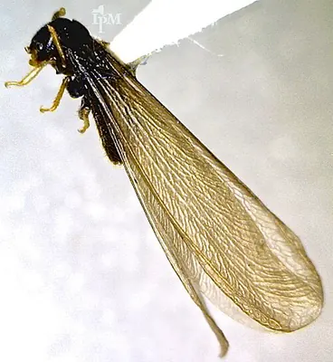
Mounted termite swarmer, species not specified. Photo: NY State IPM Program at Cornell University from New York, USA. CC BY 2.0.
Mounted termite swarmer, species not specified.
The Cornell source identifies a winged termite without a species or collection location. This mounted specimen has overlapping wings and partly obscured body and antennae; it illustrates a swarmer's overall wing outline, not a complete waist, antenna or wing-count key. Do not infer a termite species, local distribution or infestation extent from it.
Original source (new tab) · CC BY 2.0
Changes: Resized and encoded as WebP; full source composition and substantive visual content preserved. No crop, recoloring, retouching or generated diagnostic content added. Downloaded a Wikimedia-generated display-size copy because the original CDN requested standard thumbnail sizes; downloaded-source dimensions and hash are recorded separately. Derivative image licence: CC BY 2.0.
Juvenile German cockroach, dorsal view
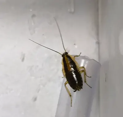
Juvenile German cockroach, dorsal view. Photo: Misei sen. CC BY-SA 4.0.
Juvenile German cockroach, dorsal view.
One juvenile, identified as a young German cockroach in the source's Japanese caption. Different instars and closely similar Blattella species need further examination; no scale is present. This photograph cannot measure infestation severity, establish a building's breeding source or identify a skin reaction.
Original source (new tab) · CC BY-SA 4.0
Changes: Resized and encoded as WebP; full source composition and substantive visual content preserved. No crop, recoloring, retouching or generated diagnostic content added. Derivative image licence: CC BY-SA 4.0.
Silverleaf whitefly adults on a leaf
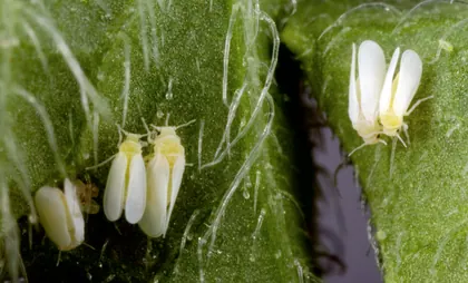
Silverleaf whitefly adults on a leaf. Photo: Stephen Ausmus. Public domain (U.S. government work). Use does not imply government agency endorsement.
Silverleaf whitefly adults on a leaf.
One whitefly species and adult stage. The plant is unspecified in the source; other whiteflies and immature stages differ. This photograph does not establish a treatment threshold.
Original source (new tab) · Public domain (U.S. government work)
Changes: Resized and encoded as WebP. Full source composition retained; no crop, retouching or changes to diagnostic features. Derivative image licence: Public domain (U.S. government work).
Citrus mealybugs, magnified laboratory sample
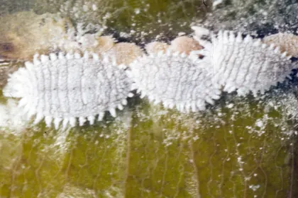
Citrus mealybugs, magnified laboratory sample. Photo: Jeffrey W. Lotz, Florida Department of Agriculture and Consumer Services, Bugwood.org. CC BY 3.0 US.
Citrus mealybugs, magnified laboratory sample.
A laboratory sample of one species. Wax and body outlines overlap, and the host plant is unspecified. Do not infer the species of a live houseplant colony from white wax alone.
Original source (new tab) · CC BY 3.0 US
Changes: Resized and encoded as WebP. Full source composition retained; no crop, retouching or changes to diagnostic features. Derivative image licence: CC BY 3.0 US.
Brown soft scale and honeydew on Ficus
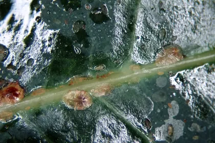
Brown soft scale and honeydew on Ficus. Photo: Whitney Cranshaw, Colorado State University, United States, Bugwood.org. CC BY 3.0 US.
Brown soft scale and honeydew on Ficus.
One soft-scale species on Ficus, with several life stages. Armoured scales and other soft scales differ; a brown bump without a visible insect is not enough to diagnose scale.
Original source (new tab) · CC BY 3.0 US
Changes: Resized and encoded as WebP. Full source composition retained; no crop, retouching or changes to diagnostic features. Derivative image licence: CC BY 3.0 US.
Western flower thrips on a leaf
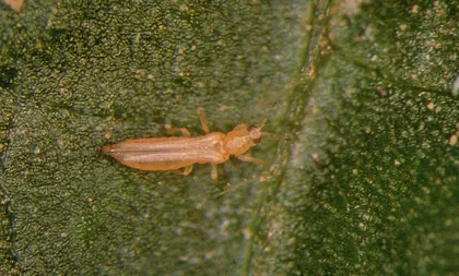
Western flower thrips on a leaf. Photo: Frank Peairs, Colorado State University, Bugwood.org. CC BY 3.0.
Western flower thrips on a leaf.
Magnified winged specimen; species identification requires finer examination than a garden photograph. The plant is unspecified and the image does not distinguish feeding damage from other causes.
Original source (new tab) · CC BY 3.0
Changes: Resized and encoded as WebP. Full source composition retained; no crop, retouching or changes to diagnostic features. Acquired the standard-size full-composition derivative offered on the source page. Commons file history records an earlier 6% vertical crop; Pest Guides made no further crop. Derivative image licence: CC BY 3.0.
Red-form female two-spotted spider mite, magnified
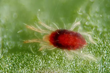
Red-form female two-spotted spider mite, magnified. Photo: Gilles San Martin from Namur, Belgium. CC BY-SA 2.0.
Red-form female two-spotted spider mite, magnified.
A focus-stacked microscope photograph of the red form, not the familiar pale form with two dark patches. The source gives body length about 0.5 mm; screen size is not a measuring scale.
Original source (new tab) · CC BY-SA 2.0
Changes: Resized and encoded as WebP. Full source composition retained; no crop, retouching or changes to diagnostic features. Acquired the standard-size full-composition derivative offered on the source page. Derivative image licence: CC BY-SA 2.0.
Spider-mite example on Cotoneaster foliage
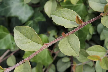
Spider-mite example on Cotoneaster foliage. Photo: I.Sáček, senior. CC0 1.0.
Spider-mite example on Cotoneaster foliage.
The source identifies Tetranychus urticae on Cotoneaster dammeri. Individual mites are not resolved here; pale leaves alone cannot separate mites from nutrient, water or other plant problems.
Original source (new tab) · CC0 1.0
Changes: Resized and encoded as WebP. Full source composition retained; no crop, retouching or changes to diagnostic features. Acquired the standard-size full-composition derivative offered on the source page. Derivative image licence: CC0 1.0.
Leaf mines attributed to Liriomyza commelinae
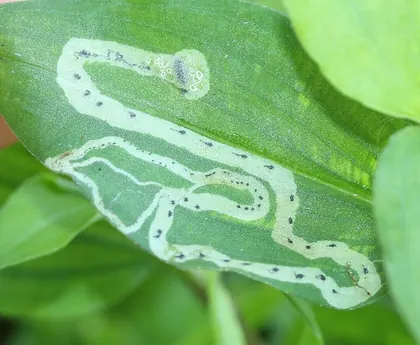
Leaf mines attributed to Liriomyza commelinae. Photo: Even Dankowicz. CC BY 4.0.
Leaf mines attributed to Liriomyza commelinae.
Shows pale winding mines and dark material within them. One leaf-miner example; mine shape alone cannot identify every miner or prove that larvae are still active. Host plant not named in the source.
Original source (new tab) · CC BY 4.0
Changes: Resized and encoded as WebP. Full source composition retained; no crop, retouching or changes to diagnostic features. Acquired the standard-size full-composition derivative offered on the source page. Derivative image licence: CC BY 4.0.
Adult tarnished plant bug in Pennsylvania
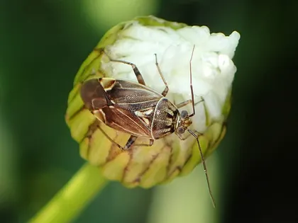
Adult tarnished plant bug in Pennsylvania. Photo: Kaldari. CC0 1.0.
Adult tarnished plant bug in Pennsylvania.
One adult plant bug. Leafhoppers, psyllids, predatory bugs and immature stages have different features. A plant bug on a plant does not by itself prove it caused the damage.
Original source (new tab) · CC0 1.0
Changes: Resized and encoded as WebP. Full source composition retained; no crop, retouching or changes to diagnostic features. Acquired the standard-size full-composition derivative offered on the source page. Derivative image licence: CC0 1.0.
Adult Japanese beetle on a flower
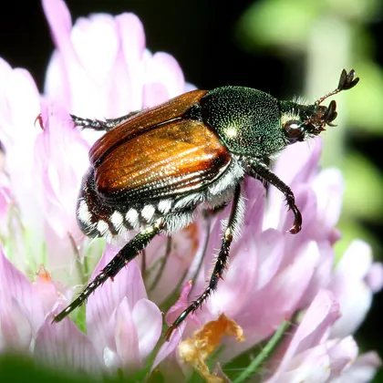
Adult Japanese beetle on a flower. Photo: Bruce Marlin. CC BY 3.0.
Adult Japanese beetle on a flower.
One adult with metallic green and copper colours and white hair tufts along the abdomen. Not a plate for all leaf beetles or weevils, and no local invasive status can be inferred from the image.
Original source (new tab) · CC BY 3.0
Changes: Resized and encoded as WebP. Full source composition retained; no crop, retouching or changes to diagnostic features. Acquired the standard-size full-composition derivative offered on the source page. Derivative image licence: CC BY 3.0.
Striped flea beetles beside feeding holes
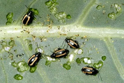
Striped flea beetles beside feeding holes. Photo: James K. Lindsey. CC BY-SA 2.5.
Striped flea beetles beside feeding holes.
Source identifies Phyllotreta vittula, photographed in Belgium. Other flea beetles differ, and the unnamed leaf is not evidence of the host range of every flea beetle. Holes alone cannot identify the cause.
Original source (new tab) · CC BY-SA 2.5
Changes: Resized and encoded as WebP. Full source composition retained; no crop, retouching or changes to diagnostic features. Derivative image licence: CC BY-SA 2.5.
Spanish slug photographed on a path
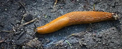
Spanish slug photographed on a path. Photo: hedera.baltica. CC BY-SA 2.0.
Spanish slug photographed on a path.
An Arion vulgaris example according to source provenance. Similar Arion species and hybrids can be difficult to separate externally; colour alone does not identify a garden slug.
Original source (new tab) · CC BY-SA 2.0
Changes: Resized and encoded as WebP. Full source composition retained; no crop, retouching or changes to diagnostic features. Acquired the standard-size full-composition derivative offered on the source page. Derivative image licence: CC BY-SA 2.0.
Brown garden snail photographed in Lima, Peru
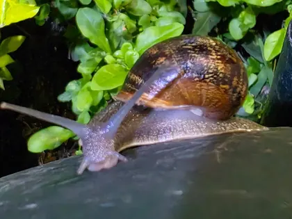
Brown garden snail photographed in Lima, Peru. Photo: Levadura5. CC BY 4.0.
Brown garden snail photographed in Lima, Peru.
A single source-labelled Cornu aspersum, with shell, head and tentacles visible. Other snails have different shells; this is not a native-status, edibility or pest-status identification.
Original source (new tab) · CC BY 4.0
Changes: Resized and encoded as WebP. Full source composition retained; no crop, retouching or changes to diagnostic features. Acquired the standard-size full-composition derivative offered on the source page. Derivative image licence: CC BY 4.0.
Codling moth larva on a cut fruit surface
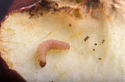
Codling moth larva on a cut fruit surface. Photo: Peggy Greb. Public domain (U.S. government work). Use does not imply government agency endorsement.
Codling moth larva on a cut fruit surface.
A magnified example of one fruit-feeding caterpillar. Maggots and other moth larvae need separate identification; the larva alone does not set the correct monitoring or treatment timing.
Original source (new tab) · Public domain (U.S. government work)
Changes: Resized and encoded as WebP. Full source composition retained; no crop, retouching or changes to diagnostic features. Acquired the standard-size full-composition derivative offered on the source page. Derivative image licence: Public domain (U.S. government work).
Galleries attributed to emerald ash borer
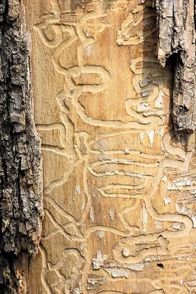
Galleries attributed to emerald ash borer. Photo: ImagePerson. CC BY 4.0.
Galleries attributed to emerald ash borer.
The photographer attributes these galleries to emerald ash borer but does not identify the tree species. Galleries alone cannot establish that species; confirm the host and insect evidence. Do not peel healthy bark to copy this view.
Original source (new tab) · CC BY 4.0
Changes: Resized and encoded as WebP. Full source composition retained; no crop, retouching or changes to diagnostic features. Acquired the standard-size full-composition derivative offered on the source page. Derivative image licence: CC BY 4.0.
Japanese beetle larva, pupae and newly emerged adults
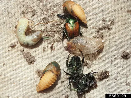
Japanese beetle larva, pupae and newly emerged adults. Photo: Whitney Cranshaw, Colorado State University, Bugwood.org. CC BY 3.0 US.
Japanese beetle larva, pupae and newly emerged adults.
A staged comparison of one beetle species; the C-shaped white larva is at upper left. Other grubs and decomposers differ. Correct grub identification and root-zone sampling matter before deciding to treat a lawn.
Original source (new tab) · CC BY 3.0 US
Changes: Resized and encoded as WebP. Full source composition retained; no crop, retouching or changes to diagnostic features. Acquired the standard-size full-composition derivative offered on the source page. Derivative image licence: CC BY 3.0 US.
Root-knot galls and egg masses on tomato roots
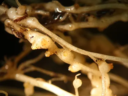
Root-knot galls and egg masses on tomato roots. Photo: Scot Nelson. CC0 1.0.
Root-knot galls and egg masses on tomato roots.
The source identifies Meloidogyne incognita on tomato. The nematodes themselves are microscopic; this photo cannot identify a nematode species or distinguish every cause of swollen roots without host history and laboratory examination.
Original source (new tab) · CC0 1.0
Changes: Resized and encoded as WebP. Full source composition retained; no crop, retouching or changes to diagnostic features. Acquired the standard-size full-composition derivative offered on the source page. Derivative image licence: CC0 1.0.
Early-blight lesions on a tomato leaf
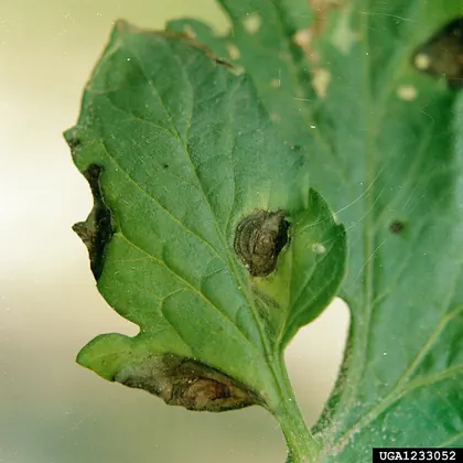
Early-blight lesions on a tomato leaf. Photo: Clemson University - USDA Cooperative Extension Slide Series. CC BY 3.0 US.
Early-blight lesions on a tomato leaf.
The source identifies Alternaria solani and target-shaped lesions on tomato. One disease example, not a diagnosis from a brown spot. Other pathogens and environmental injury can overlap.
Original source (new tab) · CC BY 3.0 US
Changes: Resized and encoded as WebP. Full source composition retained; no crop, retouching or changes to diagnostic features. Acquired the standard-size full-composition derivative offered on the source page. Derivative image licence: CC BY 3.0 US.
Powdery mildew on a squash leaf
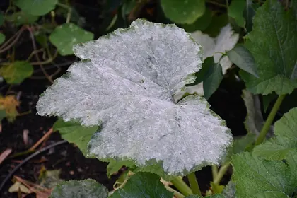
Powdery mildew on a squash leaf. Photo: Dmitry Brant. CC BY-SA 4.0.
Powdery mildew on a squash leaf.
Powdery mildew on squash according to the photographer. The fungus species is not identified. Natural leaf patterns, residues and other leaf diseases need separate consideration.
Original source (new tab) · CC BY-SA 4.0
Changes: Resized and encoded as WebP. Full source composition retained; no crop, retouching or changes to diagnostic features. Acquired the standard-size full-composition derivative offered on the source page. Derivative image licence: CC BY-SA 4.0.
Dandelion rosette with flower stalks
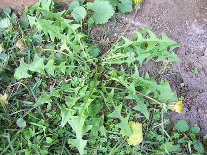
Dandelion rosette with flower stalks. Photo: Harry Rose from South West Rocks, Australia. CC BY 2.0.
Dandelion rosette with flower stalks.
One broadleaf perennial among other small plants. Leaf shape alone cannot separate every dandelion lookalike. The photograph is not an edibility guide or a reason that a plant must be removed.
Original source (new tab) · CC BY 2.0
Changes: Resized and encoded as WebP. Full source composition retained; no crop, retouching or changes to diagnostic features. Acquired the standard-size full-composition derivative offered on the source page. Derivative image licence: CC BY 2.0.
Hairy crabgrass growing along the ground
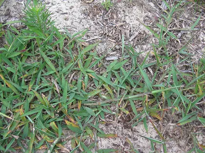
Hairy crabgrass growing along the ground. Photo: Harry Rose from South West Rocks, Australia. CC BY 2.0.
Hairy crabgrass growing along the ground.
A whole-plant example from Australia, with surrounding vegetation. Ligule, sheath hair and seedhead details are not resolved well enough here to separate every similar grass.
Original source (new tab) · CC BY 2.0
Changes: Resized and encoded as WebP. Full source composition retained; no crop, retouching or changes to diagnostic features. Acquired the standard-size full-composition derivative offered on the source page. Derivative image licence: CC BY 2.0.
Asian lady beetle larva on a leaf
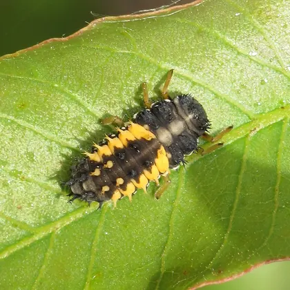
Asian lady beetle larva on a leaf. Photo: Robert Flogaus-Faust. CC BY 4.0.
Asian lady beetle larva on a leaf.
A predatory lady beetle larva, not a plant-eating caterpillar. Other lady beetle larvae vary in pattern. Asian lady beetles can also become indoor nuisance visitors; beneficial role and local invasive status are separate questions.
Original source (new tab) · CC BY 4.0
Changes: Resized and encoded as WebP. Full source composition retained; no crop, retouching or changes to diagnostic features. Acquired the standard-size full-composition derivative offered on the source page. Derivative image licence: CC BY 4.0.
Tobacco hornworm with its rear horn visible
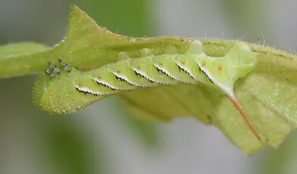
Tobacco hornworm with its rear horn visible. Photo: Calibas. CC BY-SA 4.0.
Tobacco hornworm with its rear horn visible.
The source identifies Manduca sexta. The orange-red rear horn is at the right of this photograph; diagonal side stripes and body form are visible. Other hornworms differ, and a hornworm image does not identify every cause of missing tomato foliage. The host plant is unspecified in the source.
Original source (new tab) · CC BY-SA 4.0
Changes: Resized and encoded as WebP. Full source composition retained; no crop, retouching or changes to diagnostic features. Acquired the standard-size full-composition derivative offered on the source page. Derivative image licence: CC BY-SA 4.0.
Parasitoid-wasp cocoons on a tobacco hornworm
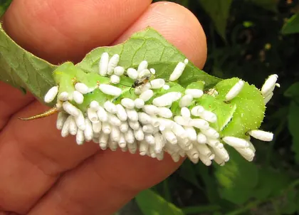
Parasitoid-wasp cocoons on a tobacco hornworm. Photo: Beatriz Moisset. CC BY-SA 4.0.
Parasitoid-wasp cocoons on a tobacco hornworm.
White structures are wasp cocoons, not caterpillar eggs. The source identifies Cotesia congregata on Manduca sexta. This example is not a plate for every parasitoid; do not handle an unknown caterpillar to reproduce the view.
Original source (new tab) · CC BY-SA 4.0
Changes: Resized and encoded as WebP. Full source composition retained; no crop, retouching or changes to diagnostic features. Acquired the standard-size full-composition derivative offered on the source page. Derivative image licence: CC BY-SA 4.0.
Adult spotted lanternfly in Pennsylvania
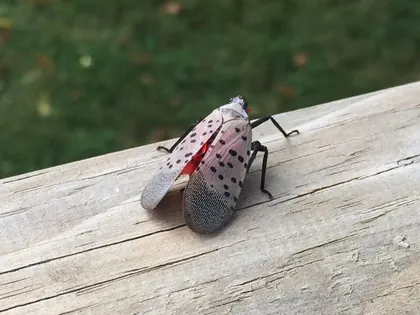
Adult spotted lanternfly in Pennsylvania. Photo: Walthery at English Wikipedia. CC BY-SA 3.0.
Adult spotted lanternfly in Pennsylvania.
One adult with spotted forewings and net-like darker tips. Red hindwings are mostly covered here. Nymphs and egg masses look different; this image does not establish local invasive status or a reporting obligation.
Original source (new tab) · CC BY-SA 3.0
Changes: Resized and encoded as WebP. Full source composition retained; no crop, retouching or changes to diagnostic features. Acquired the standard-size full-composition derivative offered on the source page. Derivative image licence: CC BY-SA 3.0.
Late-stage spotted lanternfly nymph in Pittsburgh
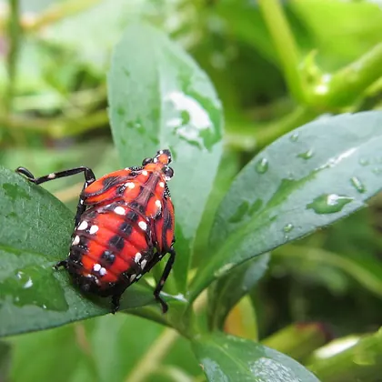
Late-stage spotted lanternfly nymph in Pittsburgh. Photo: Cbaile19. CC0 1.0.
Late-stage spotted lanternfly nymph in Pittsburgh.
Shows a red, black and white-spotted late-stage nymph. Earlier nymphs are black with white spots, while adults have wings. Do not infer regional status or management requirements from this photograph.
Original source (new tab) · CC0 1.0
Changes: Resized and encoded as WebP. Full source composition retained; no crop, retouching or changes to diagnostic features. Acquired the standard-size full-composition derivative offered on the source page. Derivative image licence: CC0 1.0.
Eastern grey squirrel in St James’s Park, London
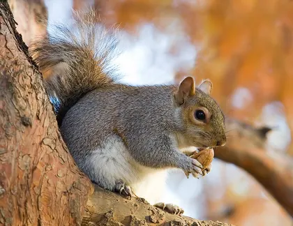
Eastern grey squirrel in St James’s Park, London. Photo: Diliff. CC BY-SA 3.0.
Eastern grey squirrel in St James’s Park, London.
One tree squirrel in a feeding pose. The tail is partly hidden; this photograph does not identify chipmunks, flying squirrels or a concealed attic occupant.
Original source (new tab) · CC BY-SA 3.0
Changes: Resized and encoded as WebP. Full source composition retained; no crop, retouching or changes to diagnostic features. Acquired the standard-size full-composition derivative offered on the source page. The Commons source is the photographer’s edited version identified in its filename. Derivative image licence: CC BY-SA 3.0.
Eastern mole above ground
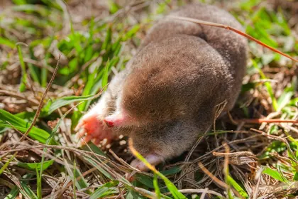
Eastern mole above ground. Photo: Bert Cash from Brunswick , USA. CC BY 2.0.
Eastern mole above ground.
Shows a pointed snout and broad digging forefeet. One mole species only; no burrow or lawn damage is shown, and a mole sighting does not identify every nearby tunnel.
Original source (new tab) · CC BY 2.0
Changes: Resized and encoded as WebP. Full source composition retained; no crop, retouching or changes to diagnostic features. Acquired the standard-size full-composition derivative offered on the source page. Derivative image licence: CC BY 2.0.
Eastern meadow vole emerging through snow
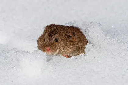
Eastern meadow vole emerging through snow. Photo: Chuck Homler d/b/a Focus On Wildlife. CC BY-SA 4.0.
Eastern meadow vole emerging through snow.
A living vole with a blunt face and small ears partly concealed by fur. The tail and most of the body are hidden, so this is not a tail-length comparison or a plate for all vole species.
Original source (new tab) · CC BY-SA 4.0
Changes: Resized and encoded as WebP. Full source composition retained; no crop, retouching or changes to diagnostic features. Acquired the standard-size full-composition derivative offered on the source page. Derivative image licence: CC BY-SA 4.0.
Botta’s pocket gopher in grass
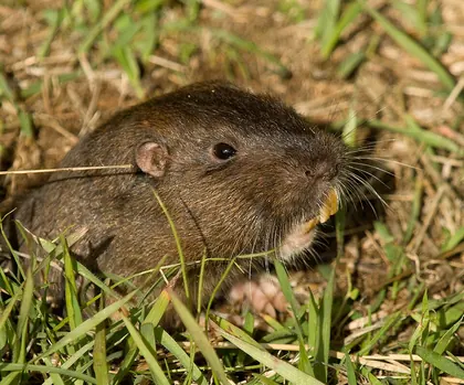
Botta’s pocket gopher in grass. Photo: Chuck Abbe. CC BY 2.0.
Botta’s pocket gopher in grass.
Shows a source-identified pocket gopher with small ears, prominent incisors and forefeet. One pocket-gopher species only; the image does not establish what made a nearby mound or burrow.
Original source (new tab) · CC BY 2.0
Changes: Resized and encoded as WebP. Full source composition retained; no crop, retouching or changes to diagnostic features. Acquired the standard-size full-composition derivative offered on the source page. Derivative image licence: CC BY 2.0.
Earth along a European mole tunnel in a Hereford garden
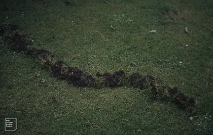
Earth along a European mole tunnel in a Hereford garden. Photo: Dr Mary Gillham Archive Project. CC BY 2.0.
Earth along a European mole tunnel in a Hereford garden.
The archive labels this as a European mole tunnel at Abbey Dore Gardens, Hereford, England. A source-labelled sign example, not an independent identification of an unknown hole or evidence for a specific North American mole species. Other animals can disturb turf.
Original source (new tab) · CC BY 2.0
Changes: Resized and encoded as WebP. Full source composition retained; no crop, retouching or changes to diagnostic features. Acquired the standard-size full-composition derivative offered on the source page. Derivative image licence: CC BY 2.0.
Raccoon standing in a garden
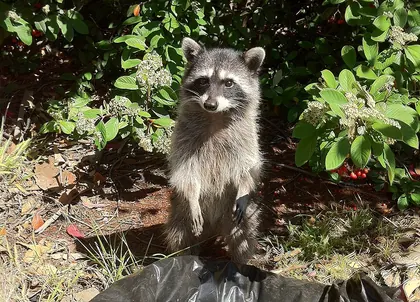
Raccoon standing in a garden. Photo: Saaws. CC BY-SA 3.0.
Raccoon standing in a garden.
Face mask and front paws are visible; the tail is hidden. The source describes an animal expecting food. Do not feed or approach wildlife to obtain an identification view.
Original source (new tab) · CC BY-SA 3.0
Changes: Resized and encoded as WebP. Full source composition retained; no crop, retouching or changes to diagnostic features. Acquired the standard-size full-composition derivative offered on the source page. Derivative image licence: CC BY-SA 3.0.
Striped skunks beside a log
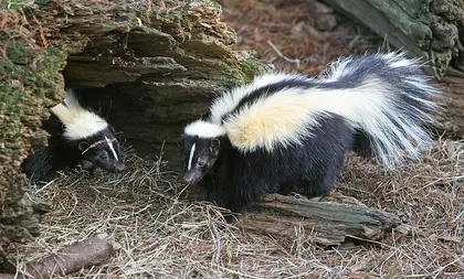
Striped skunks beside a log. Photo: Tom Friedel / BirdPhotos.com. CC BY 3.0.
Striped skunks beside a log.
Representative striped-skunk pattern; striped-skunk colour patterns vary and spotted skunks differ. A daytime sighting or a photograph cannot establish illness.
Original source (new tab) · CC BY 3.0
Changes: Resized and encoded as WebP. Full source composition retained; no crop, retouching or changes to diagnostic features. Acquired the standard-size full-composition derivative offered on the source page. Derivative image licence: CC BY 3.0.
Virginia opossum photographed at night
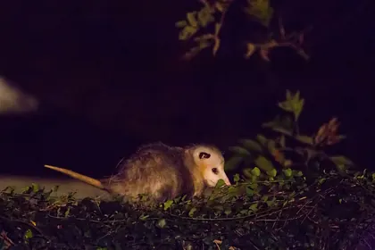
Virginia opossum photographed at night. Photo: Melissa McMasters from Memphis, TN, United States. CC BY 2.0.
Virginia opossum photographed at night.
Shows the pale face and largely bare tail, with much of the frame in darkness. One species only. The image cannot establish health, behaviour or that an animal caused nearby damage.
Original source (new tab) · CC BY 2.0
Changes: Resized and encoded as WebP. Full source composition retained; no crop, retouching or changes to diagnostic features. Acquired the standard-size full-composition derivative offered on the source page. Derivative image licence: CC BY 2.0.
Little brown bat in a house eave in Pennsylvania
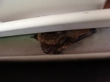
Little brown bat in a house eave in Pennsylvania. Photo: Bearerofthecup. Public domain (copyright-holder dedication).
Little brown bat in a house eave in Pennsylvania.
The source identifies a little brown bat, but a resting bat in a crevice cannot reliably be identified to species from this view. No photo determines rabies status or whether a human exposure occurred. Do not approach or disturb a roost to obtain this view.
Original source (new tab) · Public domain (copyright-holder dedication)

Changes: Resized and encoded as WebP. Full source composition retained; no crop, retouching or changes to diagnostic features. Acquired the standard-size full-composition derivative offered on the source page. Derivative image licence: Public domain (copyright-holder dedication).
Little brown bat held by a USFWS biologist
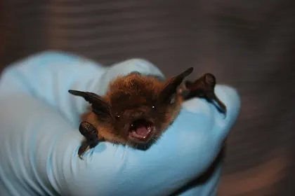
Little brown bat held by a USFWS biologist. Photo: Ann Froschauer / U.S. Fish and Wildlife Service. CC BY 2.0. Use does not imply government agency endorsement.
Little brown bat held by a USFWS biologist.
A documented research-handling scene with gloves, not a handling instruction for residents. One species only; no photograph determines disease status or replaces exposure advice.
Original source (new tab) · CC BY 2.0
Changes: Resized and encoded as WebP. Full source composition retained; no crop, retouching or changes to diagnostic features. Acquired the standard-size full-composition derivative offered on the source page. Derivative image licence: CC BY 2.0.
Feral pigeons on a tree in Chemnitz, Germany
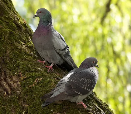
Feral pigeons on a tree in Chemnitz, Germany. Photo: Jörg Hempel. CC BY-SA 3.0 DE.
Feral pigeons on a tree in Chemnitz, Germany.
Two feral pigeons with different plumage patterns. Other pigeons, doves and building-nesting birds differ. Species protection and nest rules depend on the location; a photo is not permission to remove a nest.
Original source (new tab) · CC BY-SA 3.0 DE
Changes: Resized and encoded as WebP. Full source composition retained; no crop, retouching or changes to diagnostic features. Acquired the standard-size full-composition derivative offered on the source page. Derivative image licence: CC BY-SA 3.0 DE.
Common starling at Chester Zoo, England
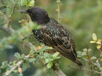
Common starling at Chester Zoo, England. Photo: Hans Hillewaert. CC BY-SA 3.0.
Common starling at Chester Zoo, England.
One starling showing dark plumage and pale feather edges. Plumage and bill colour change with age and season. Native status and legal protection differ by region.
Original source (new tab) · CC BY-SA 3.0
Changes: Resized and encoded as WebP. Full source composition retained; no crop, retouching or changes to diagnostic features. Acquired the standard-size full-composition derivative offered on the source page. Derivative image licence: CC BY-SA 3.0.
Eastern garter snake in Wooster, Ohio
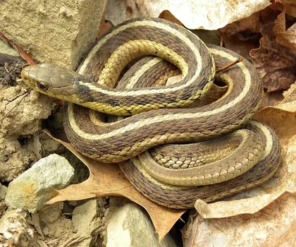
Eastern garter snake in Wooster, Ohio. Photo: Mark A. Wilson (Wilson44691). Public domain (copyright-holder dedication).
Eastern garter snake in Wooster, Ohio.
A source-identified garter snake with lengthwise stripes. Many snakes have stripes or similar colours; do not use this single example or head shape to classify an unknown snake as safe to handle.
Original source (new tab) · Public domain (copyright-holder dedication)

Changes: Resized and encoded as WebP. Full source composition retained; no crop, retouching or changes to diagnostic features. Acquired the standard-size full-composition derivative offered on the source page. Derivative image licence: Public domain (copyright-holder dedication).
Eastern fence lizard in South Carolina
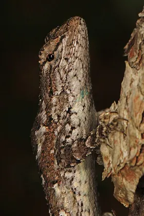
Eastern fence lizard in South Carolina. Photo: Judy Gallagher. CC BY 2.0.
Eastern fence lizard in South Carolina.
Close view of the head and scaled body, with the limbs and tail outside the frame. Not a full-body plate for all lizards or a safe-handling instruction.
Original source (new tab) · CC BY 2.0
Changes: Resized and encoded as WebP. Full source composition retained; no crop, retouching or changes to diagnostic features. Acquired the standard-size full-composition derivative offered on the source page. Derivative image licence: CC BY 2.0.
Adult white-tailed deer buck in vegetation
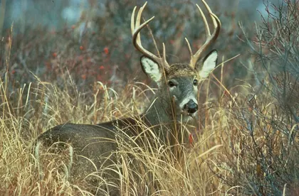
Adult white-tailed deer buck in vegetation. Photo: U.S. Fish and Wildlife Service. Public domain (U.S. government work). Use does not imply government agency endorsement.
Adult white-tailed deer buck in vegetation.
An adult male deer; females and juveniles lack the same antlers, and the tail is hidden. No browsing injury is shown, so identify plant damage independently.
Original source (new tab) · Public domain (U.S. government work)
Changes: Resized and encoded as WebP. Full source composition retained; no crop, retouching or changes to diagnostic features. Acquired the standard-size full-composition derivative offered on the source page. Derivative image licence: Public domain (U.S. government work).
Eastern cottontail sitting in grass
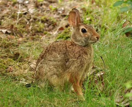
Eastern cottontail sitting in grass. Photo: The High Fin Sperm Whale. CC BY-SA 3.0.
Eastern cottontail sitting in grass.
One rabbit species with body and ears visible. Hares and other rabbits differ; this photo cannot identify the cause of a clipped stem without other evidence.
Original source (new tab) · CC BY-SA 3.0
Changes: Resized and encoded as WebP. Full source composition retained; no crop, retouching or changes to diagnostic features. Acquired the standard-size full-composition derivative offered on the source page. Derivative image licence: CC BY-SA 3.0.
Living roof rat with its long tail visible
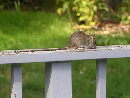
Living roof rat with its long tail visible. Photo: Alex O'Neal. CC BY-SA 2.0.
Living roof rat with its long tail visible.
The source identifies Rattus rattus; its tail extends left across the railing. Perspective and posture affect proportions, so use several traits and never image size alone to distinguish rat species.
Original source (new tab) · CC BY-SA 2.0
Changes: Resized and encoded as WebP. Full source composition retained; no crop, retouching or changes to diagnostic features. Acquired the standard-size full-composition derivative offered on the source page. Derivative image licence: CC BY-SA 2.0.
Norway rat beside a wooden wall in Lithuania
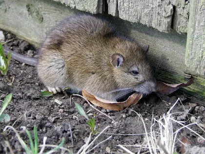
Norway rat beside a wooden wall in Lithuania. Photo: Tocekas. CC BY-SA 4.0.
Norway rat beside a wooden wall in Lithuania.
A living Norway rat according to source provenance, with the robust body, head and ear profile visible. The tail tip leaves the left edge, so this photograph cannot support a tail-to-body length measurement.
Original source (new tab) · CC BY-SA 4.0
Changes: Resized and encoded as WebP. Full source composition retained; no crop, retouching or changes to diagnostic features. Derivative image licence: CC BY-SA 4.0.
Young Norway rat in Western Washington State
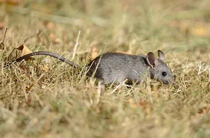
Young Norway rat in Western Washington State. Photo: VJAnderson. CC BY-SA 4.0.
Young Norway rat in Western Washington State.
The photographer explicitly labels this animal an infant Norway rat. Young rats can resemble mice and have different proportions from adults. Do not use this juvenile as an adult Norway-versus-roof-rat plate.
Original source (new tab) · CC BY-SA 4.0
Changes: Resized and encoded as WebP. Full source composition retained; no crop, retouching or changes to diagnostic features. Acquired the standard-size full-composition derivative offered on the source page. Derivative image licence: CC BY-SA 4.0.
Deer mouse photographed in a tree
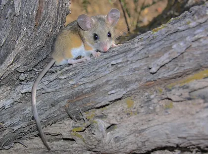
Deer mouse photographed in a tree. Photo: Gregory "Slobirdr" Smith. CC BY-SA 2.0.
Deer mouse photographed in a tree.
White feet and underside contrast with the upper fur. The historical source identifies Peromyscus maniculatus; similar Peromyscus species need finer examination and taxonomy has changed. No photograph determines infection status.
Original source (new tab) · CC BY-SA 2.0
Changes: Resized and encoded as WebP. Full source composition retained; no crop, retouching or changes to diagnostic features. Acquired the standard-size full-composition derivative offered on the source page. Derivative image licence: CC BY-SA 2.0.
Dry tomato-hornworm frass beneath an infested plant
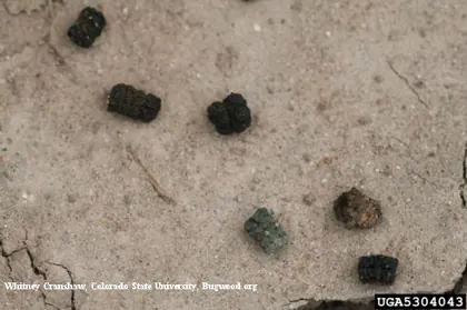
Dry tomato-hornworm frass beneath an infested plant. Photo: Whitney Cranshaw, Colorado State University, Bugwood.org. CC BY 3.0 US.
Dry tomato-hornworm frass beneath an infested plant.
The source identifies these dry pellets as frass of tomato hornworm, Manduca quinquemaculata, photographed in Colorado. They are not labelled as tobacco-hornworm frass. Droppings prompt a search for a caterpillar and fresh feeding injury; pellets alone cannot confirm the species, show that a caterpillar is still present or establish a treatment threshold.
Original source (new tab) · CC BY 3.0 US
Changes: Resized and encoded as WebP. Full source composition retained; no crop, retouching or changes to diagnostic features. Derivative image licence: CC BY 3.0 US.
Sleeping bat between boardwalk planks in Yellowstone
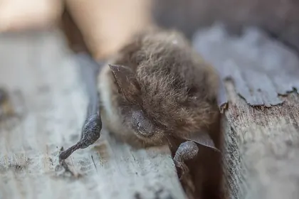
Sleeping bat between boardwalk planks in Yellowstone. Photo: Neal Herbert / National Park Service. Public domain (U.S. government work). Use does not imply government agency endorsement.
Sleeping bat between boardwalk planks in Yellowstone.
The NPS title identifies a Myotis species; its description identifies a little brown bat. The ear, fur and folded wing are visible, while most of the body is concealed. This is a resting bat photographed without handling, not a complete species-identification plate. A photograph cannot determine disease status or whether a human exposure occurred. Do not approach or disturb a bat to obtain this view.
Original source (new tab) · Public domain (U.S. government work)
Changes: Resized and encoded as WebP. Full source composition retained; no crop, retouching or changes to diagnostic features. Acquired the standard-size full-composition derivative offered on the source page. Derivative image licence: Public domain (U.S. government work).
Indianmeal moth with its two-tone folded wings visible
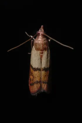
Indianmeal moth with its two-tone folded wings visible. Photo: Adrian Tync. CC BY-SA 4.0.
Indianmeal moth with its two-tone folded wings visible.
A source-identified adult on a television screen in Bytom, Poland. Pale basal forewings and copper outer sections are visible; larvae and other pantry moth species look different. Screen colour and lighting alter apparent colours, and an adult sighting does not identify the infested food.
Original source (new tab) · CC BY-SA 4.0
Changes: Resized and encoded as WebP. Full source composition retained; no crop, retouching or changes to diagnostic features. Derivative image licence: CC BY-SA 4.0.
Indianmeal moth adults among grain kernels
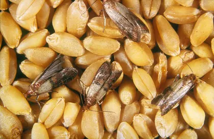
Indianmeal moth adults among grain kernels. Photo: Entomology, CSIRO. CC BY 3.0.
Indianmeal moth adults among grain kernels.
Source identifies Indianmeal moth adults. The central adult shows the pale basal and darker outer wing areas; other adults are angled or partly outside the frame. Grain in this image does not identify the food source of a particular household infestation.
Original source (new tab) · CC BY 3.0
Changes: Resized and encoded as WebP. Full source composition retained; no crop, retouching or changes to diagnostic features. Acquired the standard-size full-composition derivative offered on the source page. Derivative image licence: CC BY 3.0.
Botta’s pocket gopher at a soil opening in southern Nevada
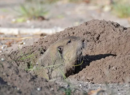
Botta’s pocket gopher at a soil opening in southern Nevada. Photo: VJAnderson. CC BY-SA 4.0.
Botta’s pocket gopher at a soil opening in southern Nevada.
The source identifies the visible animal as Botta’s pocket gopher. Loose soil and an opening are visible beside it, providing context that an unidentified hole cannot. The low angle and frame edges hide the full mound outline; this does not show a complete crescent-shaped mound from above or its usual plugged state after the animal withdraws.
Original source (new tab) · CC BY-SA 4.0
Changes: Resized and encoded as WebP. Full source composition retained; no crop, retouching or changes to diagnostic features. Acquired the standard-size full-composition derivative offered on the source page. Derivative image licence: CC BY-SA 4.0.
The inspection-house illustration
AI-generated for Pest Guides with OpenAI ImageGen on 15 October 2026; resized and compressed for the web. It shows inspection settings only. No organism, construction detail or treatment in this illustration should be used as diagnostic or engineering evidence.